
Fig. 6.
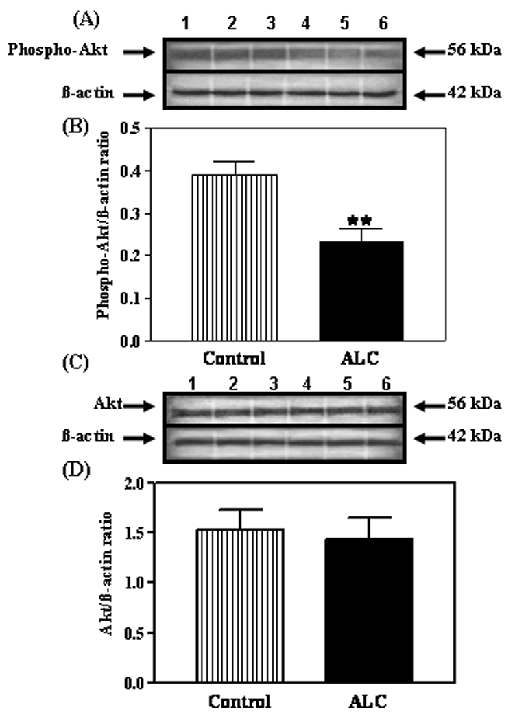
Effect of chronic ALC exposure on phosphorylated and total Akt protein expression in the ARC nucleus of prepubertal female rats. (A) Representative Western immunoblot of phosphorylated Akt and β- actin proteins isolated from control (lanes 1–3) and ALC-treated (lanes 4–6) animals. (B) Densitometric quantitation of all of the bands from two blots assessing the phosphorylated Akt protein. These data were normalized to the internal control β -actin protein, and the densitometric units represent the phosphorylated Akt/β-actin ratio. Note that chronic ALC-treated animals showed a marked decrease in phosphorylated Akt protein expression compared with control animals. (C) Representative Western immunoblot of total, non-phosphorylated Akt β-actin proteins isolated from control (lanes 1–3) and ALC-treated (lanes 4–6) animals. (D) Densitometric quantitation of all of the bands from two blots corresponding to the total, non-phosphorylated Akt protein. These data were normalized to the internal control β-actin protein, and the densitometric units represent the total Akt/β -actin ratio. Note that chronic ALC-exposure did not affect total Akt protein expression. The respective bars illustrate the mean (±SEM) of an N of 7 per group. **p<0.01 versus control.