Fig. 3.
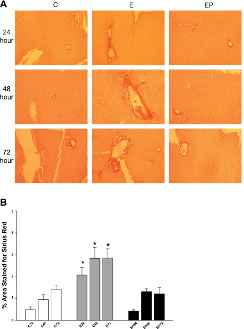
A: Sirius red stain of PCLS. PCLS were incubated in the absence (C) or presence of 25 mM ethanol (E) or ethanol + 0.5 mM 4-MP (EP) up to 72 h. At each point, PLCS were rinsed, fixed with buffered formalin, and embedded in paraffin. Sections (4 μm thick) were stained with Sirius red. Slides were analyzed with a Nikon Eclipse 80i at 10× power and Nis-Elements 3.0 software (Nikon, Melville, NY). Photographs are representative of 4 separate animal experiments. B: quantification of Sirius red staining of PCLS. PCLS were incubated in the absence or presence of 25 mM ethanol or ethanol + 0.5 mM 4-MP up to 72 h. Samples were stained for Sirius red and photographed. Pictures were deconvoluted, subjected to threshold analysis, and quantified by use of MBF-ImageJ software. PCLS slides from 4 separate animals using 10 random fields per slide were analyzed. *P < 0.01 compared with control or ethanol + 4-MP.

