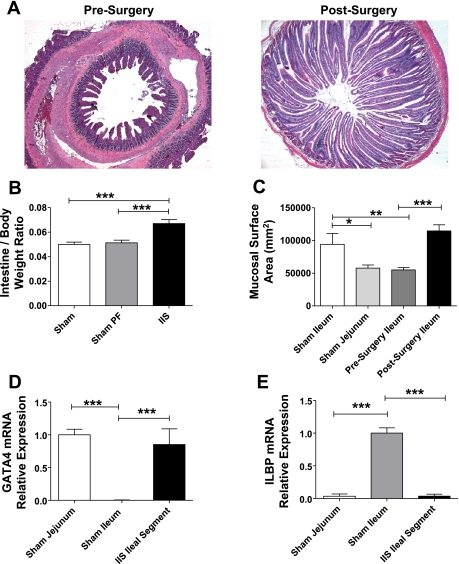
Fig. 2.
A: histological adaptation of the interposed segment. Photomicrographs (×10 magnification) of hematoxylin-and-eosin-stained sections of the same segment of ileum first at the time of surgery and then at the time of death 6 wk later. There is marked increase in the length and number of villi in the IIS rats. B: intestine-to-body weight ratios. Intestine weight normalized for body weight of the rat in IIS rats postsurgery was increased in IIS rats. For groups IIS, SH, and SH-PF, n = 6, 7, and 8, respectively (***P < 0.001). C: surface area of jejunum and ileum at surgery and study completion. Surface area of intestinal segments was estimated by using a formula of a cylinder and the ileal segment adapted to its new proximal environment to increase its surface area within 6 wk to jejunal levels. For groups IIS, SH, and SH-PF, n = 6, 7, and 8, respectively (*P < 0.05; **P < 0.01; ***P < 0.001). D: adaptation of epithelial jejunal specific gene GATA4 mRNA levels of proximal intestinal marker GATA4 were measured by RT-PCR and expressed in relative expression units to GAPDH (***P < 0.001). For groups IIS and SH, n =7 and 8, respectively. E: adaptation of epithelial ileal specific gene ILBP. mRNA levels of distal intestinal marker ILBP were measured by RT-PCR and expressed in relative expression units to GAPDH. (***P < 0.001). For groups IIS and SH, n = 7 and 8, respectively.

