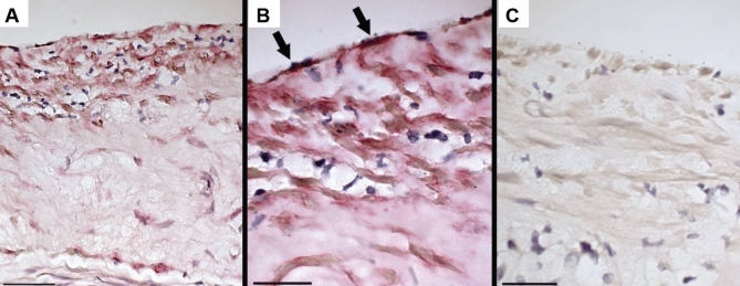
Figure 5).
Immunostaining of control atherosclerotic plaques for type IV collagen. A Low magnification shows staining in the upper regions of the plaque; scale bar = 50 μm. B High magnification shows staining for type IV collagen (arrows) in the material subtending the endothelium; scale bar = 25 μm. C Immunoglobulin G control; scale bar = 25 μm

