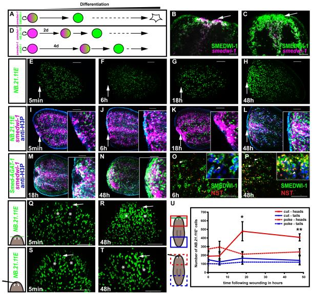
Fig. 6. The second mitotic peak is accompanied by differentiation and cell growth at the wound site.
(A) Lineage relationship schematic. smedwi-1+(mRNA, magenta)/SMEDWI-1+(protein, green) neoblasts will become SMEDWI-1+ cells as they differentiate. (B-C) Tail fragments probed with anti-SMEDWI-1 antibody (green) and smedwi-1 riboprobe (magenta). White arrow, wound site. Anterior, up. (B) smedwi-1+/SMEDWI-1+ cells accumulate at the wound site of tail fragments at 18h. White arrow, wound site. Anterior, left. (C) A layer of smedwi-1−/SMEDWI-1+ cells formed in front of actively proliferating smedwi-1+/SMEDWI-1+ cells at 48h, indicating increased differentiation. (D) smedwi-1+ neoblasts (magenta) will turn on the NB.21.11E and Smed-AGAT-1 genes (green) as they differentiate. (E-L) Timecourse, following amputation, of tail fragments (E-H) labeled with an NB.21.11E riboprobe (green); (I-L) merge of (E-H) labeling with anti-H3P antibody for mitoses (blue epidermal fluorescence is non-specific), and with a smedwi-1 riboprobe (magenta). Insets, magnified view of wound site. White arrow, wound site. Anterior, left. (M-N) Timecourse, following amputation, of tail fragments. Smed-AGAT-1+ (EC616230) cells (green) accumulate at the wound site in front of smedwi-1+ cells (magenta) at 48h (mitoses in blue, anti-H3P), indicating increased differentiation. Anterior, left. (O-P) Neoblast descendants at the wound site show an increase in nucleolar size at 48h. Tail fragments labeled with anti-SMEDWI-1 (green) and anti-NST (red) antibodies. NST signal was also found in SMEDWI-1+ cells, white arrowheads. (O) At 6h, NST was localized to a small nuclear region, inset (Hoechst, blue). (P) At 48h, NST signal occupies up to 1/3 of the nucleus. SMEDWI-1 and NST are in the cytoplasm and nucleus, respectively. However, both antibodies are from rabbits, leading to some artificial double labeling where there was high signal intensity (e.g., at 48h, nucleolar SMEDWI-1 signal is an artifact). (Q-S) Increased neoblast descendant formation at wounds is specific to tissue loss. NB.21.11E+cells (green) increased at the wound site (white arrow) between 18h and 48h following head tip amputation (Q-R), but not following a poke in the head tip (S-T). (U) Number of NB.21.11E+ cells in the wound area shown in images (Q-T). Data represent averages; n ≥ 3 ± sd. Green line, amputation plane. Red and blue regions, analyzed areas. (B, C, E-T) Images represent superimposed optical sections, dorsal view, unless otherwise stated. Scale bars, 100μm.

