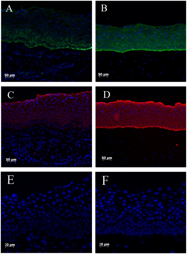
Figure 1. K14 and K3 expression in limbal and central corneal regions.
K14 (Green) positive cells were only detected to the basal layer of limbus (A), but its expression was also seen throughout central corneal layers (B). Although K3 (Red) expression is absent in the limbal basal layer cells (C), K3 highlights whole central corneal cell layers (D). Control sections for limbal (E) and central cornea (F) show no background staining.

