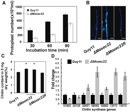
Figure 8. Deletion of MoSEC22 alters the fungal cell wall.
(A) Protoplasts released under the treatment of cell-wall-degrading enzymes. The released protoplast was quantified at 30 min intervals. Data comprise three independent experiments, with triple replications each time yielding similar results. (B) Disruption of MoSEC22 (#1) altered the distribution of chitin on the cell wall. The experiment was repeated several times with triple replications that yielded similar results. (C) GlcNa determination by the fluorimetric Morgan–Elson method shows increased chitin contents in the ΔMosec22 mutant. Asterisks indicate a significant difference between the sporulation in the mutant and wild-type strains (or the reconstituted strain) at p = 0.01, according to Duncan's range test. Data comprise three independent experiments with triple replications each time that yielded similar results. (D) Transcription analysis of seven M. oryzae chitin synthases using qRT-PCR.

