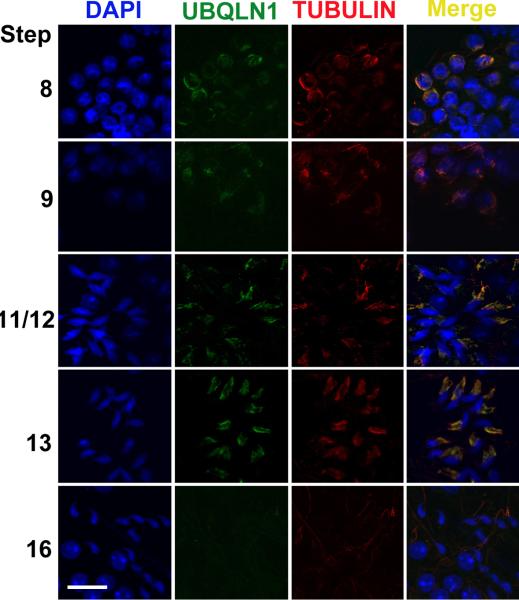
Fig. 5.
Immunofluorescent localization of UBQLN1 to the manchette of elongating spermatids. Green fluorescence represents the UBQLN1 immunoreactivity and red fluorescence indicates the immunoreactivity of β-Tubulin, a marker for the manchette. Cell nuclei were counterstained using DAPI. All panels are in the same magnification. Scale bar = 10μm.

