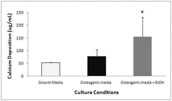
Figure 5. The effect of ethanol on calcium deposition.

AFSCs were exposed to 100 mM ethanol during the first 48 hours of osteogenic differentiation. At day 23 of differentiation, AFSCs were stained for calcium deposition using alizarin red staining. AFSCs exposed for 48 hours to 100 mM ethanol had significantly increased calcium deposition when compared to non-exposed AFSCs (155.1 ± 75.8 μg/mL vs 77.4 ± 26.9 μg/mL). Calcium deposition was detected in non-differentiated AFSCs at a basal level of 53.3 ± 2.2 μg/ml. The values shown are the mean +/− SD (n=3–6) and similar results were confirmed in another cell line (data not shown). * One-tailed t-test of significance of p<0.006.
