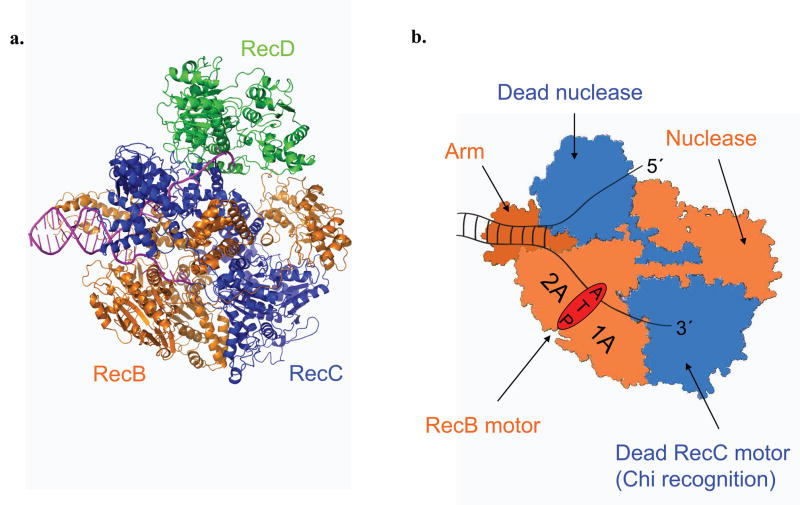
Figure 1. RecBCD and RecBC structures.
RecB (orange), RecC (blue), and RecD (green) subunits are indicated. (a). Ribbon diagram of a RecBCD–DNA complex 6,21. (b). Cartoon depiction of a RecBC–DNA complex. RecB motor, nuclease, and arm domains are indicated along with the catalytically dead RecC motor and nuclease domains. The paths of the 3′- and 5′-terminated unwound ssDNA are shown.

