
Figure 3. The secretin architecture is conserved in different secretion systems.
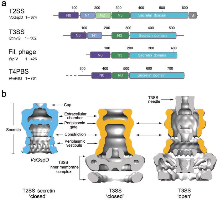
(a) Domain architecture of secretins from the T2SS, the T3SS, the filamentous phage assembly system and the T4 pilus biogenesis system (T4PBS). Members of the secretin super-family contain a C-terminal secretin core homology domain (cyan)17,18. The T2SS secretins generally contain four periplasmic subdomains, termed N0-N3. The N0 subdomain (dark blue) is located at the N-terminus and is followed by the three structurally homologous subdomains, N1-N3 (blue, green and dark green, respectively). A T2SS specific domain, termed the S-domain (grey), is located at the very C-terminus. Secretins from other systems share a similar architecture, composed of the secretin domain and at least two periplasmic subdomains that are structurally equivalent to N0 and N3 of VcGspD. (b) Structural comparison of the VcGspD density (blue, left) to single particle reconstructions of the T3SS in its close state (center) (EMDB 122426) and to the fully assembled T3SS needle complex in its open state (right) (EMDB 161724). The outer membrane T3SS secretin sits on top of a large inner membrane complex (gold, center and right). VcGspD appears to be in its closed state (left compared with center).