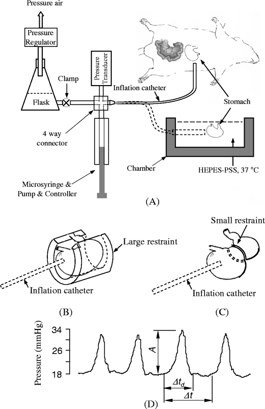
Fig. 1.

a A schematics of isovolumic myograph and in vivo and ex vivo experimental setup. The four-way connector merges the inflation flask, pressure transducer, microsyringe, and extended tubing. The chamber contains the HEPES-PSS at 37°C. The stomach and myograph are at isovolumic state when clamp is closed. b A large restraint limited gastric distension. c A small restraint applied on the stomach. d Typical pressure waves of gastric contraction. A is amplitude, Δt d is the duration of contraction, Δt is period
