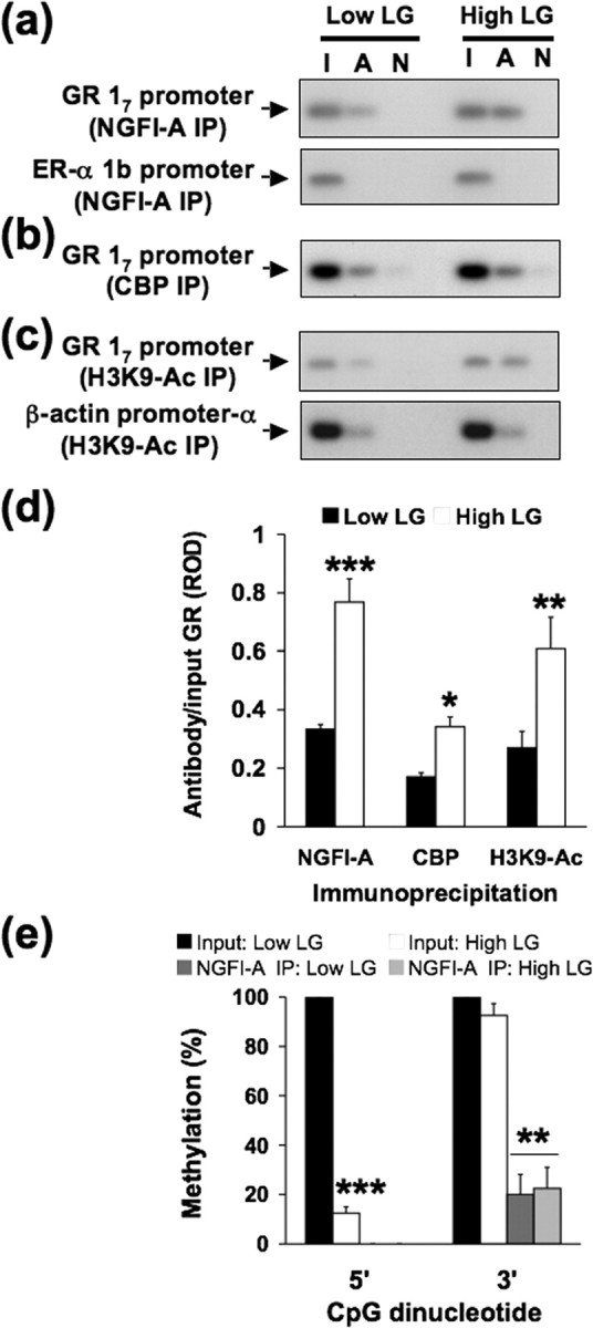
Figure 7.

Chromatin immunoprecipitation of CBP, histone H3–K9 acetylation and NGFI-A binding to the exon 17 GR and DNA methylation analysis of NGFI-A-bound exon 17 GR sequence in hippocampal tissue of 6-d-old high and low LG offspring (n = 4 animals per group). a–c, Lanes were loaded with non-immunoprecipitated input (I), CBP, acetylated histone H3–K9 or NGFI-A primary antibody immunoprecipitated (A), or non-immune IgG antibody immunoprecipitated (N) hippocampal extracts. a, Representative Southern blots of the amplified exon 17 region of the GR from NGFI-A immunoprecipitated (IP) hippocampal tissue (194 bp band). Exon 1b estrogen receptor-α promoter region, which does not contain NGFI-A recognition elements (493 bp), amplified from the same NGFI-A immunoprecipitated hippocampal tissue was run as a control for specificity and showed no signal. b, Representative Southern blots of the amplified exon 17 region of the GR from CBP immunoprecipitated hippocampal tissue (194 bp band). c, Representative Southern blots of the amplified exon 17 region from acetyl-histone H3–K9 immunoprecipitated hippocampal tissue (194 bp band) and β-actin (171 bp band) control. d, ROD (mean ± SEM) of exon 17 sequence amplified from cAMP, acetyl-histone H3–K9, or NGFI-A immunoprecipitated hippocampal tissue of 6-d-old high and low LG offspring (n = 4 animals per group; *p < 0.05; **p < 0.001; ***p < 0.0001). e, Mean ± SEM percentage methylation per cytosine for the 5′ and 3′ CpG dinucleotides within the NGFI-A binding region of the exon 17 GR promoter bound to NGFI-A protein in 6-d-old offspring of high and low LG mothers (6–10 clones sequenced per animal; n = 4 animals per group; **p < 0.001; ***p < 0.0001).
