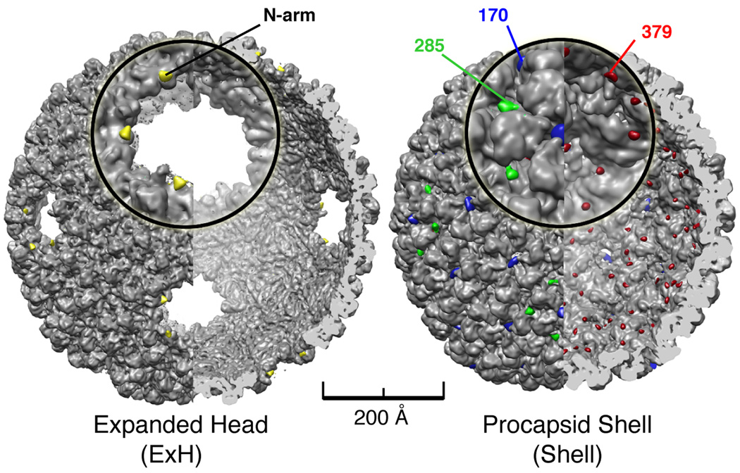
Figure 2. Identification of P22 N-arms in ExH and nano-gold labeled residues in shell mutants.
(Left) Cutaway view along two-fold direction of digested ExH reconstruction (computed to 7.0 Å, gray shaded surface) showing superimposed densities ascribed to the N-arms (yellow) obtained from an ExH minus digested ExH difference map. The ExH and digested ExH reconstructions were both computed at 14.0 Å resolution for difference map analysis. An enlarged view of one vertex appears in the insert. (Right) Same as left panel but showing that difference densities corresponding to nano-gold labels in reconstructions of three shell variants pinpoint the locations of residues 170 (blue), 285 (green), and 379 (red) in the WT procapsid shell (computed to 9.0Å, gray shaded-surface). Difference map analyses were all performed with gold-labeled and WT shell maps computed to 11.0 Å resolution.

