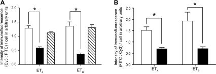
Fig. 10.
A: quantitation of the immunofluorescence intensity of ETA and ETB (CY3-labeled) receptors to FITC-labeled macrophages in control (open bars), HPAH (solid bars), and IPAH (diagonal bars) lungs. Intensity of immunofluorescence for ETA and ETB receptor antibody is significantly reduced in macrophages from HPAH lungs compared with controls and IPAH cases. BMPR2 mutant mice compared with controls. B: ETA (FITC-labeled) and ETB (FITC-labeled) receptors in macrophages (Cy3-labeled) from control (open bar) and BMPR2 mutant (solid bar) mice. Intensity of immunofluorescence for ETA and ETB receptor antibody is significantly reduced in macrophages from BMPR2 mutant mice compared with controls. *P < 0.001.

