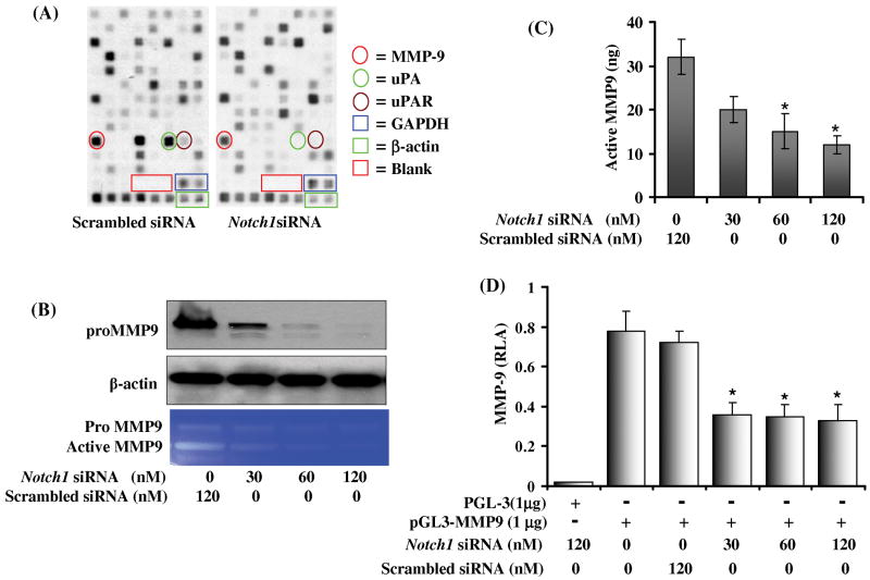
Figure 3. Effect of Notch1 knockdown in PC3 cells on the expression of genes involved in extracellular matrix degradation and cell adhesion.
(A) Autoradiographic image of a cDNA array from scrambled siRNA transfected control (Left) and Notch1 knockdown cells (Right). Enriched tetraspots indicate the position of genes. Red encircle MMP9; green encircle uPA and brown encircle uPAR. (B) Western blot analysis to evaluate pro MMP9 protein expression in Notch1 knockdown cells. Blot was reprobed with β-actin antibody to analyze the equal loading of proteins. (C) Gelatin zymogram showing activity of pro MMP9 and active MMP9 in scrambled and Notch1 siRNA transfected cells. (D) Quantification of MMP9 secretion using MMP9 specific ELISA performed in the culture media of PC3 cells transfected either with scrambled or Notch1 specific siRNA. (E) Effect of Notch1 knockdown on the promoter activity of MMP9 gene expression in PC3 cells. 2×10⁶ PC3 cells cotransfected with either scrambled or Notch1 specific siRNA along with 1 μg of pGL3 or 1μg MMP9 luciferase reporter plasmid and 50 ng of renilla luciferase reporter plasmid as an internal control as described in material and method section. The experiment was performed in quadruplet and the values are showing mean ± SD. Asterisk (*) represents p<0.01 as significant.

