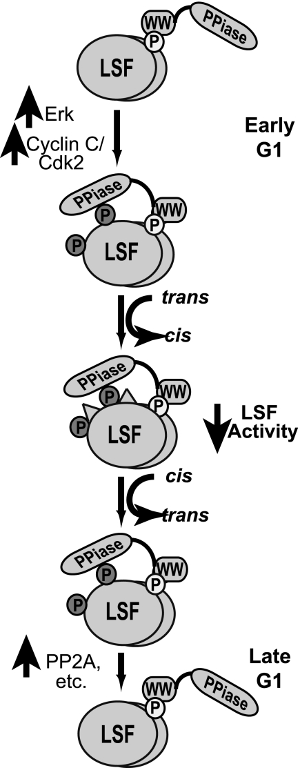
FIGURE 7.
Model of Pin1 regulation of LSF. A detailed discussion of the model is provided in the text. Only the LSF modifications discussed in this study are indicated, with their relationship to Pin1. Other protein-protein interactions and modifications that may differentially alter LSF activities are not shown or discussed here; see Ref. 7 for some additional insights. LSF is indicated as a dimer of ovals, with phosphorylation sites shown only on a single subunit, for clarity of presentation. Phosphorylated residues are indicated as small circles: Thr-329, light circle; Ser-291 and Ser-309, dark circles. The cis configuration of the Ser-Pro bonds in LSF is indicated as raised triangles on the surface of the oval. Pin1 is indicated as two domains: PPiase is the catalytic domain and WW is the binding domain; a flexible linker in the form of a line connects the two domains.

