FIGURE 6.
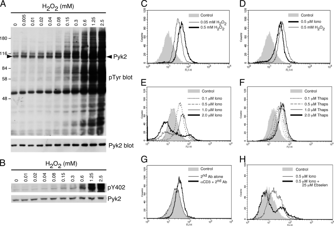
H2O2 stimulates Pyk2 phosphorylation and inducers of Pyk2 phosphorylation stimulate production of reactive oxygen species in CTL. A, AB.1 were stimulated with the indicated concentration of H2O2 for 10 min then lysed. Shown are total cell lysates probed with anti-phosphotyrosine followed by Pyk2. The arrowhead in the top blot indicates the position of Pyk2. B, as in A except cell lysates were probed sequentially with antibodies specific for pY402-Pyk2 and Pyk2. C, AB.1 cells were left as a control or stimulated with 0.05 mm or 0.5 mm H2O2 for 10 min at 37 °C and assessed for the presence of ROS after incubation for 15 min with the indicator dye H2DCFDA, as described under “Experimental Procedures.” D, AB.1 cells were treated with DMSO carrier control, 0.5 μm ionomycin or 0.5 mm H2O2 for 10 min at 37 °C and assessed for ROS. E, AB.1 cells were treated with DMSO control (corresponding to the highest amount added to the cells as carrier) or with increasing ionomycin concentrations as indicated prior to ROS assessment. F, as for E except cells were treated with various concentrations of thapsigargin. G, AB.1 cells were left untreated or treated with secondary cross-linking antibody alone or in combination with anti-CD3 and assessed for the presence of ROS. H, AB.1 cells were pretreated with or without 25 μm ebselen then stimulated with 0.5 μm ionomycin for 10 min at 37 °C prior to ROS detection. The control contains the highest amount of DMSO added to the cells. All data are representative of at least three independent experiments.

