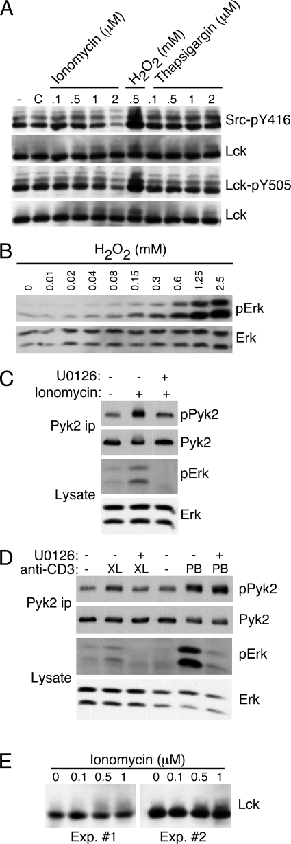
FIGURE 8.
Erk phosphorylation is stimulated by H2O2 or ionomycin and is required for optimal ionomycin-stimulated Pyk2 phosphorylation. A, AB.1 were stimulated with the indicated concentration of ionomycin, H2O2 or thapsigargin for 10 min. (−) indicates untreated cells and C indicates cells treated with DMSO carrier. Cell lysates were prepared and duplicate blots were probed sequentially with antibodies specific for Src-pY416 then Lck or Lck-pY505 followed by Lck. B, AB.1 were stimulated with the indicated concentration of H2O2 for 10 min. Cell lysates were prepared and probed sequentially with antibodies specific for phospho-Erk then Erk. C, AB.1 were stimulated with 0.5 μm ionomycin for 10 min in the presence or absence of 10 μm U0126. Cell lysates and Pyk2 immunoprecipitates were prepared and probed with the indicated antibody. D, AB.1 were stimulated with cross-linked (XL) anti-CD3 for 10 min or with plate-bound (PB) anti-CD3 for 20 min in the presence or absence of 10 μm U0126. Cell lysates and Pyk2 immunoprecipitates were prepared and probed with the indicated antibody. E, AB.1 were stimulated with the indicated concentration of ionomycin for 10 min and cell lysates probed with anti-Lck. Shown are blots from two separate experiments.

