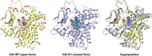
FIGURE 3.
Ribbon diagram of overall structure of Vdh-WT (open form) and Vdh-K1 (closed form). Overlay of these two structures is also shown. The heme cofactor is in the sphere, and the bound-substrate (25(OH)VD3) and the side chains of mutational residues at positions 70, 156, 216, and 384 are in sticks. The α-helices are labeled alphabetically from the N to C terminus.

