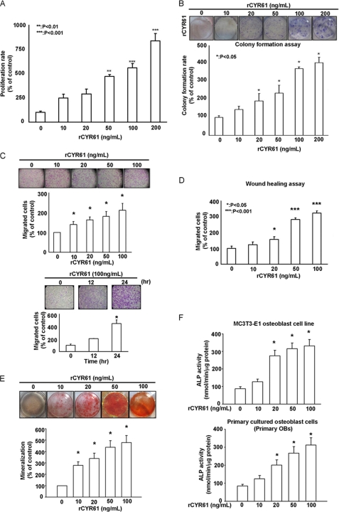
FIGURE 1.
Recombinant CYR61 protein induces proliferation, migration, and osteoblastic differentiation of MC3T3-E1 cells. A, treatment with rCYR61 increased proliferation per MTT assay is shown. After treatment at various doses, growth rates were measured by MTT assay. B, treatment with rCYR61 increased proliferation per colony counts. After 7 days colonies were stained with crystal violet and counted. C, migration ability of MC3T3-E1 cells increased after rCYR61 treatment. Cells that migrated were stained with crystal violet and counted (upper). Cells were incubated with 100 ng/ml rCYR61 in Transwell plates for indicated times. Cells that migrated were stained with crystal violet and counted (lower). D, treatment with rCYR61 increased wound-healing migration. Still images were captured at the indicated times after wounding, and then cells were counted. E, cells were incubated with rCYR61 in the indicated doses. After 14 days cells were stained with Alizarin red (upper). The quantitative data of mineralization ability are shown in the lower panel. F, ALP activity assay identified osteoblastic differentiation of MC3T3-E1 and primary cultured osteoblasts after rCYR61 treatment. Cells were incubated with rCYR61 in the indicated doses. After 14 days, cells were collected to determine ALP activity. The upper panel indicates enzyme activity of MC3T3-E1 cells; the lower panel indicates enzyme activity of primary cultured osteoblasts (OB). Each experiment was performed in triplicate, and results represent the mean ± S.D. of three independent experiments. The asterisks indicate a significant difference (*, p < 0.05; **, p < 0.01; ***, p < 0.001) between rCYR61 treatment and vehicle treatment cells.

