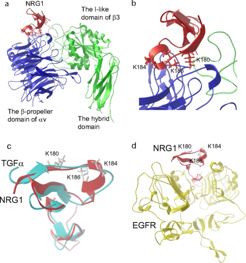
FIGURE 2.
Docking simulation of αvβ3-NRG1 interaction. a, a model of NRG1-integrin αvβ3 interaction predicted by docking simulation by using AutoDock3 is shown. The headpiece of integrin αvβ3 (PDB code 1LG5) was used as a target. The model predicts that the EGF-like domain of NRG1 (PDB code 1HAF, blue) binds to the RGD-binding site of the integrin αvβ3 headpiece (green and red). b, the Lys residues at positions 180, 184, and 186 of NRG1α are located at the interface between NRG1 and αvβ3 and were selected for mutagenesis studies. c, superposition of TGFα and NRG1 is shown. d, the Lys residues at positions 180, 184, and 186 of NRG1 are not located in the binding site for EGFR. We replaced TGFα in the TGFα-EGFR complex (PDB code 1MOX) with NRG1 (PDB code 1HAF) by superposing. ErbB3 or ErbB4 is homologous to EGFR.

