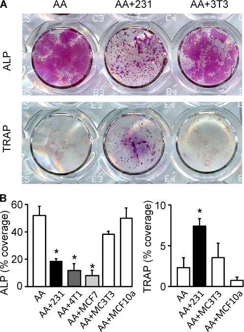
FIGURE 1.
Breast cancer cells inhibit osteoblasts and stimulate osteoclasts. Mouse bone marrow cells were grown for 3–15 days with AA (50 μg/ml) without additions (open bars) or in the presence of MDA-MB-231, 4T1, or MCF7 CM (10%, shaded bars) or controls MC3T3-E1 CM (10%) and MCF10A CM (10%). A, representative images of cultures treated with AA only (AA, left), with AA and MDA-MB-231 CM (AA+231, center), or with AA and MC3T3-E1 CM (AA+3T3, right), fixed on day 6–9, and stained for ALP (red, upper) or TRAP (purple, lower). Scanned are the wells of a 24-well plate. B, average area covered on day 9 by ALP-positive cells (left) and on day 6 by TRAP-positive cells (right). Treatment with MDA-MB-231, 4T1, or MCF7 CM significantly reduced ALP-positive osteoblast staining (left). Treatment with MDA-MB-231 CM significantly increased TRAP-positive osteoclast staining (right). Supplementation of cultures with AA and conditioned medium from MC3T3 or MCF10A did not produce significantly different results from treatment with AA alone. Data are means ± S.E. (error bars), n = 2–6 independent experiments, p < 0.05.

