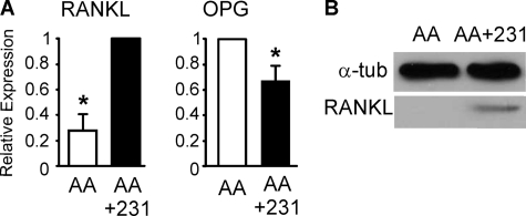
FIGURE 3.
Breast cancer cells induce osteoclastogenic change in RANKL/OPG expression. Bone marrow cells were grown for 9 days with AA (50 μg/ml) in the absence (AA, open bars) or presence of MDA-MB-231 CM (10%, AA+231, filled bars). A, expression of RANKL and OPG normalized to expression of β-actin and presented relative to levels observed in cells grown with AA+231 for RANKL and AA only for OPG. Data are means ± S.E. (error bars), n = 5 independent experiments, p < 0.05. B, RANKL protein level assessed by immunoblotting in whole cell lysates. Shown is a representative immunoblot with α-tubulin as a loading control.

