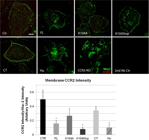
FIGURE 2.
Stimulation of microglia by N terminus-containing MCP1 proteins triggers CCR2 internalization. Primary microglia from wild-type mice activated overnight with 100 ng/ml LPS were treated with saline (Ctr) or 10 nm recombinant MCP1 proteins, rodent (FL, K104A, K104Stop, and CT) or human (Hu), for 1 h and then swelled in hypotonic buffer for 20 min to cause cell lysis. The plasma membrane sheets that remained adhered to the coverslips were stained for Mac-2 (green) to visualize the membrane sheets and CCR2 (red). CCR2−/− microglia (CCR2 KO) served as a control for the CCR2 antibody. Microglia were also immunostained with secondary antibodies (2nd Ab Ctr) only. Scale bar, 10 μm. The fluorescence intensity was quantified in 20 cells/condition in three separate experiments. The results were analyzed with one-way analysis of variance plus Dunn's test (*, p < 0.05). Error bars, S.D.

