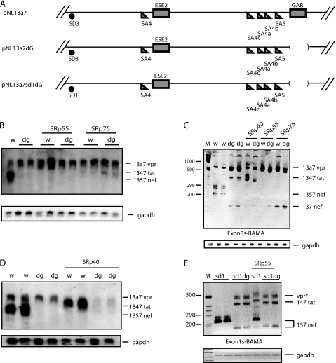
FIGURE 6.
A, schematic representation of segments of the HIV-1 subgenomic plasmids pNL13a7 (7), pNL13a7dG, and pNL13a7sd1dG. B and D, Northern blot with RNA extracted from HeLa cells transfected with pNL13a7 (w) or pNL13a7dG (dg) in the absence or presence of CMV promoter-driven plasmids expressing SRp40, SRp55, or SRp75. The blots were probed with exon 1 probe (Fig. 1A). All detected mRNAs are indicated. C, RT-PCR with RNA extracted from HeLa cells transfected with pNL13a7 (w) or pNL13a7dG (dg) in the absence or presence of CMV promoter-driven plasmids expressing SRp40, SRp55, or SRp75. RT-PCR oligonucleotides Exon3s and BAMA (Fig. 1A) were used. The bands representing mRNAs are indicated. M represents the molecular weight marker. E, RT-PCR with RNA extracted from HeLa cells transfected with pNL13a7sd1 (sd1) (Fig. 3A) or pNL13a7sd1dG (sd1dg) in the absence or presence of CMV promoter-driven plasmid expressing SRp55. Oligonucleotides used for RT-PCR were Exon1s and BAMA (Fig. 1A). Bands representing mRNAs are indicated. M represents the molecular weight marker.

