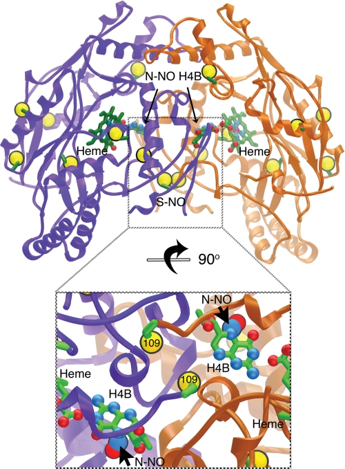
FIGURE 7.
Selective iNOSoxS- and N-nitrosation at the dimer interface (subunits colored purple/gold, cysteine sulfurs shown as large gold spheres). The nitrosylated iNOSox crystal structure (top) shows the location of our confirmed S-nitrosylation (S-NO) and N-nitrosation (N-NO-H4B) sites in the interlocked dimer interface, as well as overall distribution of cysteine residues. The inset below shows a close-up view of the S-NO and N-NO sites in the iNOSox dimer interface rotated 90° from the top image.

