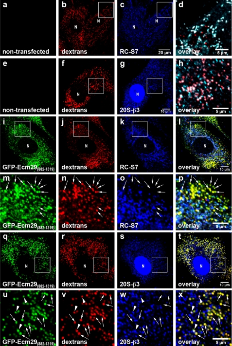
FIGURE 7.
Expression of GFP-Ecm29(882–1319) inhibits association of 26 S proteasomes with endosomes. Transfected mouse embryonic fibroblasts were grown on coverslips for 48 h prior to 1-h labeling with 1 mg/ml Alexa® 568-dextran conjugate (dextrans) and processing for confocal microscopy. 26 S proteasomes were stained with antibodies to either RC subunit S7 (RC-S7) or 20 S proteasome subunit β3 (20S-β3). Enclosed areas are magnified in panels d and h or directly below the corresponding full-size images (panels m–p and u–x). The enclosed areas in panels c and g were pseudo-colored green (not shown) and then overlaid with the corresponding red (panels b and f) and blue images (panels c and g). Therefore, dextran-filled, proteasome-bound endosomes in panels d and h are shown white rather than pink and are clearly distinguished from dextran-filled endosomes that lack proteasomes (shown in red). Note that, unlike proteasomes in non-transfected cells (panels a–h), 26 S proteasomes in transfected MEF for the most part do not localize to dextran-filled early endocytic vesicles (shown in yellow; examples are indicated by the arrows in panels m–p and u–x). The arrowheads in panels u–x indicate a few dextran-filled vesicles to which both GFP-Ecm29(882–1319) and proteasomes localize. N, cell nucleus. Scale bars, 5 or 10 μm as indicated.

