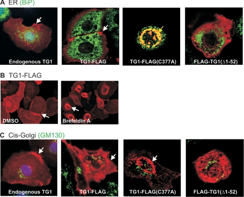
FIGURE 3.
TG1-FLAG intracellular localization. Keratinocytes were transfected with 2 μg of plasmid encoding the indicated proteins and at 48 h cells were stained with the appropriate antibody. A, TG1-FLAG(C377A) colocalizes with BiP. Cells were fixed and stained with anti-FLAG (right three images, red) or rabbit anti-TG1 (left image, red) and with anti-BiP (green). The white arrows indicate TG1 (left panel) and TG1-FLAG location (second from left panel). The yellow arrow (third from left panel) indicates the perinuclear TG1-FLAG(C377) colocalization with BiP. These are confocal 1-μm optical sections. B, TG1-FLAG accumulates in ER with brefeldin A treatment. Keratinocytes were infected with 10 m.o.i. of tAd5-TG1-FLAG and after 3 h treated with 10 μm brefeldin A for 18 h. The cells were stained with anti-FLAG for epifluorescence detection (arrows). C, wild-type and TG1 mutant proteins do not localize with GM130. Cells were incubated with anti-FLAG (right three images) or anti-TG1 (left image) (red) and with anti-GM130 (green). The white arrows indicate TG1 and TG1-FLAG membrane localization, and TG1-FLAG(C377A) perinuclear location. These are confocal 1-μm confocal images.

