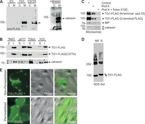
FIGURE 5.
TG1 is associated with the ER membrane. A, microsomal localization of TG1-FLAG and TG1-FLAG(C377A). Keratinocytes were infected with 10 m.o.i. of tAd5-EV, tAd5-TG1-FLAG, or tAd5-TG1-FLAG(C377A) and at 48 h total cell lysates were prepared and separated into cytosol (C) and 100,000 × g pellet (microsomal, M) fractions. Equal cell equivalents of each fraction were electrophoresed for detection of anti-FLAG and anti-calnexin. The arrow indicates migration of TG1-FLAG and TG1-FLAG(C377A). B, TG1-FLAG is associated with the ER membrane. Microsomal membranes were extracted on ice for 30 min with 1 m NaCl, 0.1 m Na2CO3 (pH 11), or 1% Triton X-100 followed by centrifugation at 100,000 × g for 1 h. The resulting soluble (S) and insoluble (I) fractions were electrophoresed for immunoblot with anti-FLAG and anti-calnexin. C, TG1-FLAG localizes inside the ER. Microsomes from tAd5-TG1-FLAG-infected cells were divided into identical aliquots and incubated with 100 μg/ml of proteinase K in the absence or presence of 1% of Triton X-100 on ice. After 30 min the samples were electrophoresed for immunoblot with anti-FLAG. D, TG1 disulfide bonds. Lysates from TG1-FLAG expressing cells were prepared in phosphate-buffered saline containing 1% Triton X-100 and boiled for 5 min in the absence (NR) or presence (R) of reducing agent. Extracts were then electrophoresed on a reducing agent-free SDS-containing 7.5% polyacrylamide gel for immunoblot with anti-FLAG. The arrows indicate migration of reduced and non-reduced monomeric TG1-FLAG. The slower migrating material (≥250 kDa) is cross-linked TG1-FLAG. E, intracellular and extracellular TG1-FLAG. At 48 h after infection with 10 m.o.i. of tAd5-TG1-FLAG, cells were fixed with 4% paraformaldehyde with or without methanol permeabilization, and incubated with anti-FLAG. Primary antibody binding was visualized using Alexa 488-conjugated goat anti-rabbit IgG secondary antibody. These are confocal 1-μm images.

