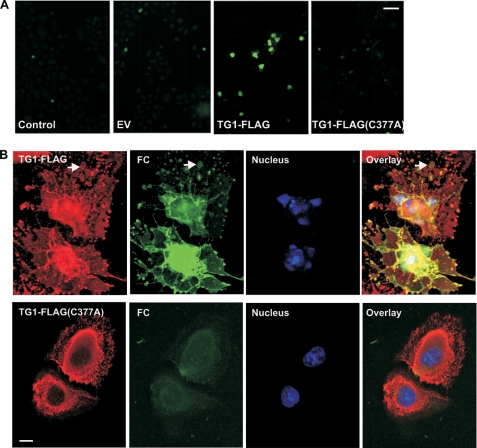
FIGURE 7.
Transglutaminase activity assay. A, cells were infected with 10 m.o.i. of tAd5-EV, tAd5-TG1-FLAG, or tAd5-TG1-FLAG(C377A) and after 48 h incubated with 100 μm FC for 4 h before detection of FC by epifluorescence. Bar = 50 μm. B, cells, treated as above, were monitored for FC fluorescence (green), TG1-FLAG (anti-FLAG, red), and the nuclear stain (Hoechst, blue). Bar = 10 μm. The signal in the TG1-FLAG(C377A) FC panel is background fluorescence. These are 1-μm confocal sections.

