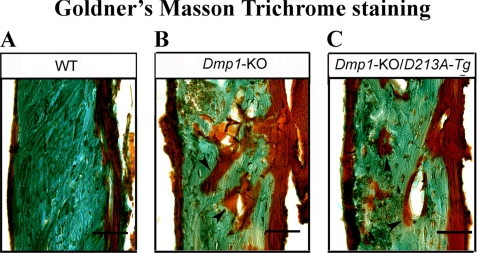
FIGURE 5.
Goldner's Masson Trichrome staining. The specimens were from the diaphysis region of the tibia of 6-week-old WT (A), Dmp1-KO (B), and Dmp1-KO/D213A-Tg (C) mice. The tibia of the Dmp1-KO and Dmp1-KO/D213A-Tg mice had remarkably more osteoid and poorly mineralized cortical bone (shown in “red”) than of the WT mice. Bars, 20 μm.

