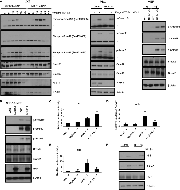
FIGURE 2.
The effects of eliminating or overexpressing NRP-1 on Smad protein phosphorylation and the Smad target gene expression. A, eliminating NRP-1 up-regulated Smad1/5 phosphorylation and down-regulated Smad2/3 phosphorylation. NRP-1 was knocked down by siRNA in LX2 and PSC. For MEF cells, wt MEF, and NRP-1−/− MEF cells were used. All the cells were serum-starved overnight, and then treated with 10 ng/ml TGF-β1 for the indicated time. An asterisk (*) indicates the nonspecific band. B, overexpress NRP-1 in NRP-1−/− MEF cells down-regulated Smad1/5 phosphorylation and up-regulated Smad2/3 phosphorylation. NRP-1−/− MEF cells were infected with NRP-1 encoding retrovirus. LacZ retrovirus was used as control. After 36 h, cells were serum-starved overnight, and then treated with 10 ng/ml TGF-β1 for the indicated time. C–E, effect of knocking down NRP-1 on the Smad proteins transcriptional activity in the cells measured by the luciferase promoter assay. 5 × 10³/well LX2 cells were plated into a 96-well plate transfected with NRP-1 siRNA for 24–30 h, then serum-starved overnight. The cells were then transfected with the corresponding luciferase promoter plasmids together with pRL-TK Renilla luciferase plasmid as the internal control. One hour after the transfection, TGF-β1 was added to the corresponding well to the final concentration of 10 ng/ml. Firefly luciferase and Renilla luciferase activities were performed. C, Luc-Id-1 promoter. D, Luc-ARE, co-transfected with FAST. E, Luc-SBE. F, knocking down NRP-1 affected the Smad protein responding gene expression. Cells were transfected with NRP-1 siRNA for 24–30 h in the complete medium, then serum-starved overnight. 10 ng/ml TGF-β1 was added to the cells for 24 h. In response to TGF-β1 stimulation, Id-1 protein (the Smad1-responding gene) expression was more highly up-regulated in NRP-1 knocked down cells than in control cells stimulated with TGF-β1. In response to TGF-β1 stimulation, α-SMA and PAI-1 protein (the Smad3-responding gene) expression was more highly down-regulated in NRP-1 knocked down cells than in control cells stimulated with TGF-β1.

