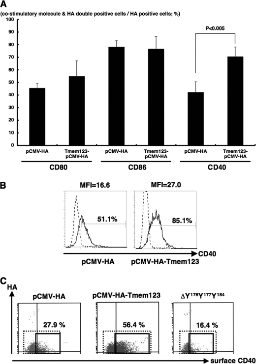
FIGURE 7.
Transfection with Tmem123 cDNA up-regulated cell surface expression of CD40 on DC2.4 cells. DC2.4 cells were transfected with pCMV-HA or pCMV-HA-Tmem123 and stained with anti-CD80 mAb, anti-CD86 mAb, and anti-CD40 mAb. These cells were permeabilized and stained with anti-Tmem123 pAb followed by phycoerythrin-conjugated goat anti-rabbit IgG Ab and analyzed by flow cytometry. A, the ratio of CD80-, CD86-, and CD40-positive cells in the HA-Tmem123- or HA-positive cells is shown. The ratio of CD40-positive cells was significantly higher in the HA-Tmem123-positive transfectants compared with HA-positive transfectants (69.9 ± 7.7 versus 42.3 ± 6.7%, n = 3, p < 0.001), although the expression levels of CD80 and CD86 did not differ. Results are mean ± S.D. (n = 3). B, the level of cell surface expression of CD40 on DC2.4 cells transfected with pCMV-HA (left panel) and pCMV-HA-Tmem123 (right panel) is shown. Solid lines, CD40 mAb; dashed lines, isotype control (representative of three independent experiments). MFI, median fluorescence intensity. C, the cell surface expression of CD40 was higher in pCMV-HA-Tmem123-transfected cells than that in mock-transfected cells. There was no difference in cell surface CD40 expression between ΔY176Y177Y184-transfected cells and mock-transfected cells. A representative of three independent experiments is shown.

