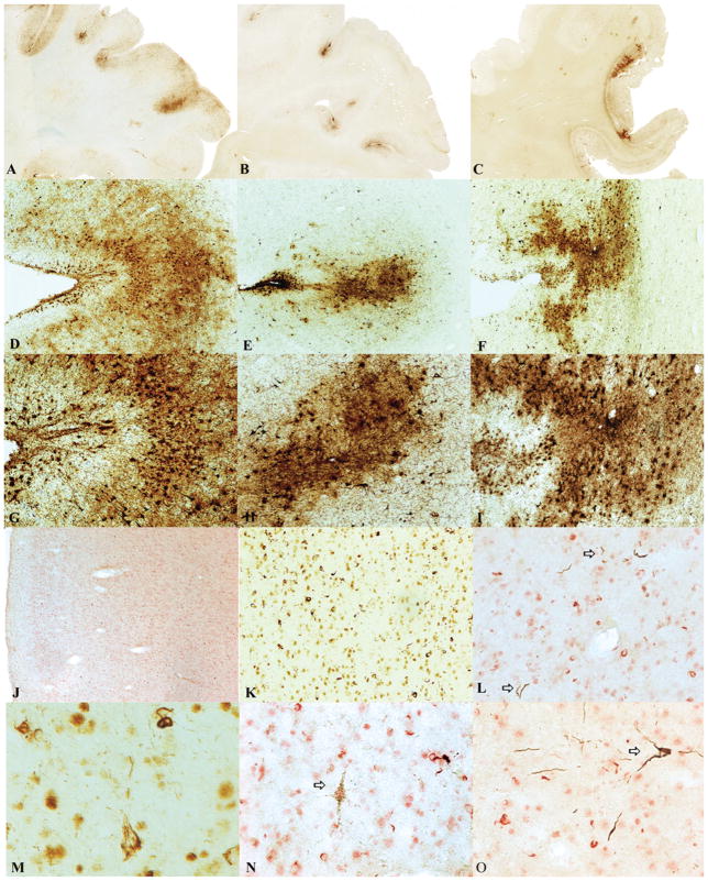
FIGURE 1.
Cases of chronic traumatic encephalopathy (CTE) with widespread TAR DNA-binding protein of approximately 43 kd (TDP-43) immunoreactivity and motor neuron disease (CTE + MND). (A–C) Tau-immunoreactive neurofibrillary degeneration in the frontal cortex of the 3 cases of CTE + MND (whole-mount 50-μm sections immunostained for CP13, original magnification: 1×). (D–I) Tau-positive neurofibrillary tangles, glial tangles, and neuropil neurites are particularly dense at the depth of the cortical sulci, CP13 immunostain, original magnification: (D–F) 50×; (G–I) 100×. (J) TDP-43 immunostaining reveals abundant TDP-positive pathology in all cortical layers in the frontal cortex of Case 1, original magnification: 50×. (K) Numerous TDP-43–positive ring-shaped neurites (RNs) and ring-shaped glial inclusions (RGIs) in the frontal cortex of Case 2, original magnification: 200×. (L) Double immunostaining shows that most TDP-43–positive RNs and RGIs (red) are not colocalized with tau-positive neurites (PHF-1 brown, arrows), original magnification: 400×. (M) TDP-43–positive filamentous neuronal inclusions, original magnification: 600×. (N) Double immunostaining shows a tau-positive pretangle (PHF-1 brown, arrow) that is not associated with TDP-43 immunoreactivity (red), original magnification: 400×. (O) Double immunostaining shows a tau-positive tangle (PHF-1, arrow) that is not associated with TDP-43 immunoreactivity (red), original magnification: 400×.

