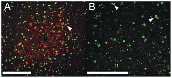
FIGURE 4.
Limited colocalization of tau and TAR DNA-binding protein of approximately 43 kd (TDP-43). (A, B) Laser scanning confocal microscopy was carried out on sections from the frontal cortex of Case 1 (A) and Case 2 (B). Sections were double immunostained for TDP-43 (green) and PHF-1 (red). Areas of colocalization appear yellow. In (A), there are abundant tau-positive neurites around a small blood vessel; white arrowhead indicates a small cluster of tau-positive neurites that seem to be colocalized with TDP-43. (B) Many TDP-43–positive neurites (green) and a few tau-positive neurites (red). Most neurites do not seem to be colocalized, but arrowheads point to occasional colocalized intraneuronal inclusions and neurites. White bars = 200 μm.

