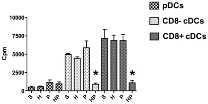
Figure 8. Mixed lymphocyte reaction assay.
Four groups of mice were studied: sham-treated (S group), hemorrhaged mice (H group), methicillin-susceptible S. aureus (MSSA)-induced pneumonia (P group), and hemorrhage before MSSA-induced pneumonia (HP group). DCs subsets were defined by specific membrane markers: B220 and siglec H for plasmacytoid DCs (pDCs), CD11c and CD8 to differentiate CD8+ conventional DCs (cDCs) from CD8− cDCs. DCs subsets were sorted and treated for 24 hours with CpG 1826 (5 µM). Each DCs subset was cultured with allogeneic CD4+ and CD8+ T cells (ratio DCs:T cells, 1∶25) for 3 days. For each DCs subset, determining thymidine incorporation within 8 hours assessed T-cell proliferation. Data are representative of three independent experiments (each group, n = 6). Histograms represent mean ± SEM. *P<0.05 versus all others.

