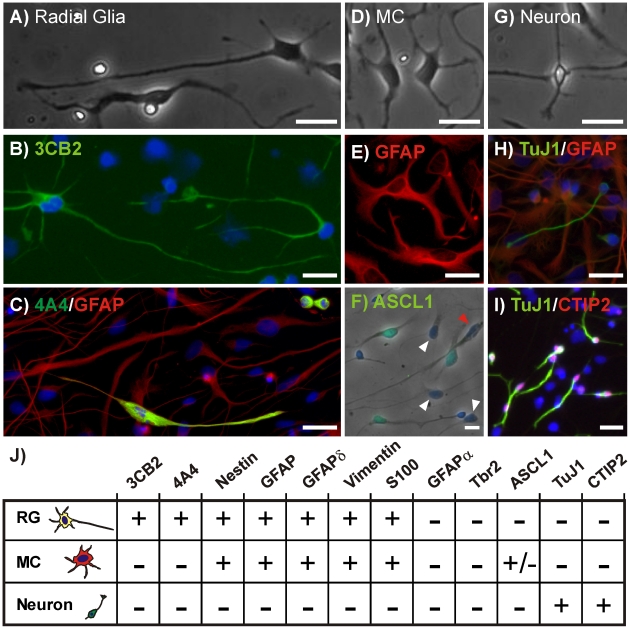
Figure 1. Three Distinct hNPC Morphologies and Marker Profiles.
hNPC-derived radial glia (RG) displayed radial morphology with processes extending over 100 um (A) and expressed the radial glial markers 3CB2 (B), 4A4 and GFAP (C). Multi-polar cells (MC) (D) stained positively for GFAP (E) and the IPC marker ASCL1 (F). ASCL1 was not expressed in all MCs (white arrowheads) nor in any of the RGs (red arrowhead). Neurons (G) stained positively for TuJ1 (H) and CTIP2 (I). Scale bar = 25 um. (J) Marker profiles for hNPC cell types. All images are representative examples of the immunocytochemistry performed for each marker protein.

