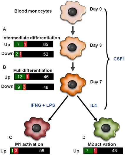
Figure 3. Graphical representation of differentially regulated genes in endometrial and blood CD14+ cells that have been reported to be differentially regulated during macrophage differentiation.
Martinez et al. (23) identified genes that are differentially expressed in monocytes after 3 (intermediate differentiation; A) or 7 days (full differentiation; B) of culture with CSF1 to cause differentiation into macrophages. Afterwards, macrophages were polarized towards the classical activation (M1) pathway [C, achieved by culture with interferon-γ (IFNG) and lipopolyssacharide (LPS)) or alternative activation (M2) pathway (D, achieved by culture with interleukin 4 (IL4)). For each stage of differentiation, bars represent the number of genes in the Martinez et al. (23) data set that were found in the present data set of endometrial and blood CD14+ cells. Genes that are upregulated in endometrium are in green, genes upregulated in blood are in red, and genes that were not significantly upregulated are in black.

