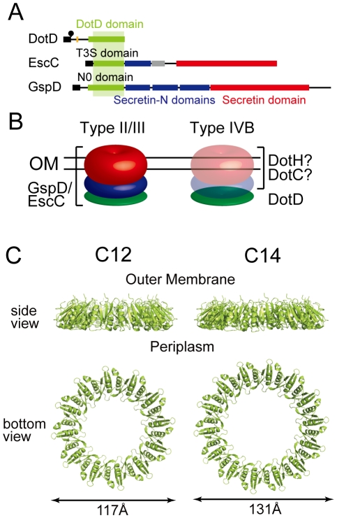
Figure 5. Periplasmic ring models of DotD.
(A) Domain organizations of DotD, EscC and GspD. Green boxes: DotD/T3S/N0 domain; Blue boxes: Secretin_N domain (protein family database Pfam PF03958); Red boxes: Secretin domain (Pfam PF00263). (B) Schematic drawings of the type II/III secretin and a putative outer membrane complex containing DotD. Red, blue and green torus represents domains schematically drawn in panel A. (C) Ring models of the DotD domain having 12- and 14-fold rotation symmetry. Ring structures were modeled using SymmDock program [47] fed with DotD atomic coordinates (excluding water molecules and the lid) and order of rotation symmetry (C12 or C14).

