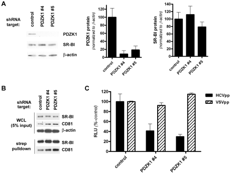
Figure 4. Stable knockdown of PDZK1 expression in HepG2(N6)+CD81 cells.
(A) HepG2(N6)+CD81 cells expressing the indicated shRNA constructs were cultured under polarizing conditions (5 d post-confluence) prior to Western analysis of PDZK1 (∼70 kDa) and SR-BI (∼85 kDa) protein levels. β-actin (∼42 kDa) was used as a loading control. PDZK1 and SR-BI protein levels were quantified by densitometry and normalized to those of the loading control β-actin (graphs). Data are means + SEM (n = 3). (B) Cell-surface expression levels of SR-BI. HepG2(N6)+CD81 cells (5 d post-confluence) expressing the indicated shRNA targets were surface-biotinylated prior to detergent lysis, streptavidin-precipitation of biotinylated proteins and Western blotting. SR-BI, PDZK1 and CD81 (∼26 kDa) protein levels in whole cell lysates (WCL) (upper panels) and streptavidin-precipitates (lower panels) are shown. Overexpressed CD81 (with a C-terminal Myc epitope tag) was detected with an anti-C-myc antibody. (C) Confluent HepG2(N6)+CD81 cells expressing the indicated shRNA constructs were infected with HCVpp or VSVpp 72 h prior to measurement of luciferase activity. Data are means + SEM (n = 4).

