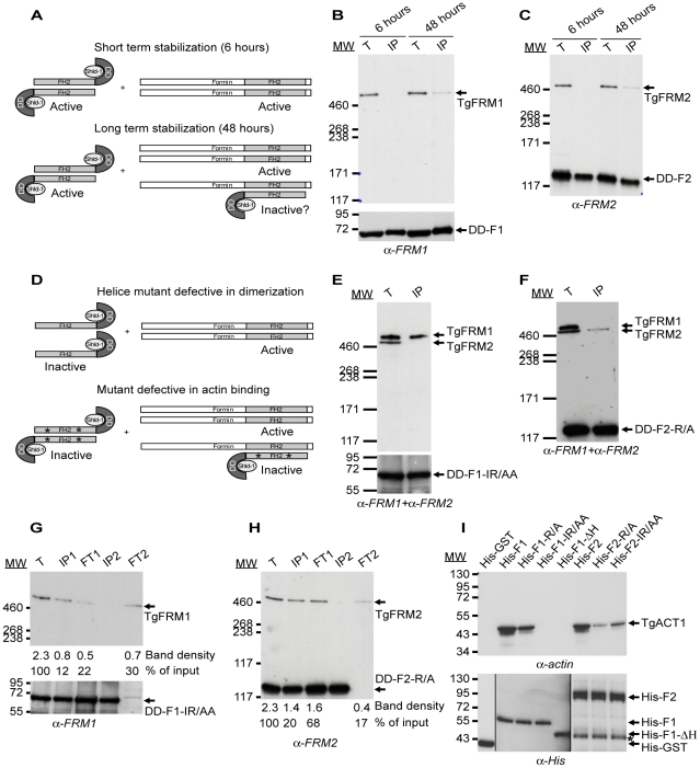
Figure 4. Expression of FH2 domains and their interaction with both endogenous formins and TgACT1 proteins.
(A) Schematic representation of the FH2 homodimers fused to DD and FH2/FRM heterodimers upon short and long-term stabilization with Shld-1. (B–C) FH2/FRM heterodimers are formed 48 hrs post stabilization of the FH2 domain. Transgenic parasites were grown in presence of Shld-1 for 6 or 48 hrs prior to harvesting of the parasites and IP with anti-myc antibodies. TgFRM1 (B) and TgFRM2 (C) were monitored by western blot using anti-FRM1 and anti-FRM2 antibodies. Parasite total lysates (T), immunoprecipitated proteins eluted from beads (IP). (D) Schematic representation of FH2-ΔH mutants fused to DD and lacking the two helices responsible for dimerization. Point's mutations represented by asterisks caused a defect in actin binding without affecting dimerization. (E–F) DD-F1-IR/AA and DD-F2-R/A formed selective heterodimers with their corresponding formin. Transgenic parasites were grown in presence of Shld-1 for 48 hrs prior to harvesting of the parasites and IP with anti-myc antibodies. Membranes were probed simultaneously with anti-FRM1 and anti-FRM2 antibodies. (G–H) Depletion of the total lysates from F1-IR/AA/FRM1, F1-IR/AA/F1-IR/AA, F2-R/A/FRM2, and F2-R/A/F2-R/A by two sequential immunoprecipitations with anti-myc antibodies. Total lysates (T) or immunoprecipitated proteins eluted from beads (IP1 and IP2) or lysates after immunoprecipitation (flow through 1 (FT1) and FT2) were analysed by Western blots. Membranes were probed with either anti-FRM1 (G) or anti-FRM2 (H) antibodies. The integrated densities of the bands measured using the ImageJ program, and the values expressed in percentage of the total input are provided after normalization for equal loading. (I) Nickel affinity pull-down assay, which measured the ability of FH2 domains fused to His to bind to TgACT1. The amount of TgACT1 and FH2 domains were determined by Western blot analysis using anti-actin and anti-His antibodies. The asterisk represents F2 domains degradation.

