Figure 1.
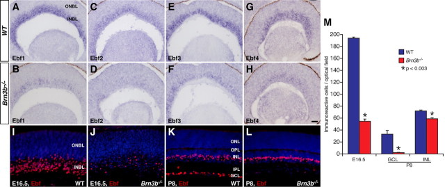
Altered expression of Ebfs in the Brn3b mutant retina. A–H, Sections from E14.5 wild-type and Brn3b−/− retinas were in situ hybridized with the indicated Ebf probes. Compared with the wild-type retina, there is a great decrease of Ebf1-4 expression in the mutant retina. I–L, Retinal sections from wild-type and Brn3b mutant mice at the indicated stages were immunostained with a pan-Ebf antibody and weakly counterstained with DAPI. In the mutant retina, there is a dramatic decrease of Ebf-immunoreactive cells within the INBL and GCL at E16.5 and P8, respectively; whereas, those within the INL at P8 show only a small reduction. M, Quantitation of Ebf-immunoreactive cells in Brn3b wild-type and mutant retinas. Each histogram represents the mean ± SD for 4 retinas. IPL, Inner plexiform layer; ONBL, outer neuroblastic layer; OPL, outer plexiform layer. Scale bar (in H): A–H, 50 μm; I–L, 25 μm.

