Fig. 4.
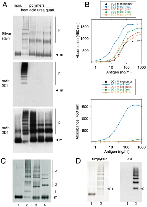
Monoclonal antibody 2C1 recognizes polymers of α1-antitrypsin prepared by heating but not by other denaturing conditions. (A) 7.5% (w/v) nondenaturing PAGE to show polymer analyzed by silver stain (upper) or Western blot, first with mAb 2C1 (center) and then with mAb 2D1 (bottom) on the same membrane. mAb 2C1 recognized heat-induced polymers but gave no signal for polymers prepared by treating α1-antitrypsin at low pH, or with 1–3 M guanidine or 1–4 M urea. The monomer (m) and polymer (p) are indicated. (B) The same polymers assayed in sandwich ELISA, using either mAb 9C5 (upper graph) or 2C1 (lower graph) as the detecting antibodies. mAb 9C5 detected all species with high affinity but mAb 2C1 detected only heat-induced polymers. (C) 7.5% (w/v) nondenaturing PAGE. Lane 1: M α1-antitrypsin monomer; lane 2: heat-induced α1-antitrypsin polymer; lane 3: antithrombin hexapeptide-induced polymer of α1-antitrypsin; lane 4: P9-P10 reactive center loop-cleaved polymer of α1-antitrypsin. The monomer (m), dimer (d), polymer (p) and intermediate state (I) are indicated. The intermediate in lane 2 migrates between the monomeric and dimeric states of the non heat-induced polymers. (D) 7.5% (w/v) nondenaturing PAGE of Z α1-antitrypsin monomer (lane 1) and polymer (lane 2) visualized by SimplyBlue™ (left) or following Western blot analysis with mAb 2C1 (right). Arrowheads indicate the intermediate (I) in the polymer lanes. The 2C1 shows no signal for the monomer but recognizes the intermediate and higher order polymers.

