Fig. 3.
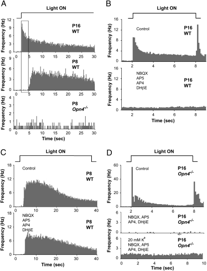
Multielectrode array (MEA) recordings of retinal ganglion cell spiking demonstrate that rod and cone-mediated light responses do not drive ipRGCs or nonipRGCs in mice younger than P10. (A) Histograms of light-evoked spikes from RGCs in response to a 40 s step of light. Top trace: P16 WT mouse; middle trace: P8 WT mouse, and bottom trace: P8 Opn4−/− mouse. (Note: although spontaneous waves of spiking were observed in P8 retinas, none of the spike activity shown here was induced by light.) Only the P16 recording (top record) shows short latency responses attributable to rod and cone signals. Top, middle, and bottom traces are averages of 16, 26, and 5 RGCs that responded to the onset of light, respectively. (B) Histograms of light-evoked spikes recorded from P16 WT mouse. Light stimuli were 6 s in duration, and the timescale is expanded compared with A. Top trace: Responses in control saline. Bottom trace: Recordings from same retina in synaptic blocker mixture of NBQX (20 μM, AMPA/KA glutamate receptor antagonist), DL-AP5 (100 μM, NMDA receptor antagonist), DL-AP4 (20 μM, mGluR6 agonist that blocks light responses in ON bipolar cells) and DHβE (2 μM, an agonist for nicotinic ACh receptors). All light-evoked spiking was eliminated in this mixture. Top and bottom traces are averages of 102 and 85 RGCs, respectively, recorded in the same retina. (C) Histograms of longer latency light-evoked spike responses from ipRGCs in P8 mouse. Top trace: control saline. Bottom trace: Synaptic blocker mixture. Same synaptic blockers used in B did not eliminate the light-evoked ipRGC responses. Top and bottom traces are averages of 66 and 62 RGCs, respectively, recorded in the same retina. Preservation of the long latency sustained light responses attributable to ipRGCs was observed in four of four retinas treated with the drug mixture (P8 and P9). (D) Histograms of light-evoked spikes in P16 Opn4−/− retina. Top trace: Short latency rod- and cone-mediated ON and OFF responses recorded in control saline. Middle trace: recordings in synaptic blocker mixture used in B and C. Bottom trace: spiking activity recorded in response to 20 mM K+ in the presence of the synaptic blocker mixture. Top, middle and bottom traces are averages of 56, 12, and 45 RGCs, respectively, recorded in the same retina. These records demonstrate that rod- and cone-mediated light responses are present in older Opn4−/− mice, and that all this activity is blocked with synaptic blockers, yet the RGCs themselves are still capable of spiking when depolarized with potassium.

