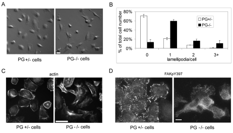
Fig. 2.
Plakoglobin regulates actin cytoskeleton organization in mouse keratinocytes. (A) DIC images of PG+/− and PG−/− cells. Scale bar: 20 μm. (B) Number of lamellipodia per cell (PG+/− white bars; PG−/− black bars). (C) Alexa-Fluor-350-conjugated phalloidin-actin staining of PG+/− and PG−/− cells. Scale bar: 20 μm. (D) PG+/− and PG−/− keratinocytes stained for FAK(Tyr397-P) (FAKpY397) to show the focal contacts. Scale bar: 20 μm.

