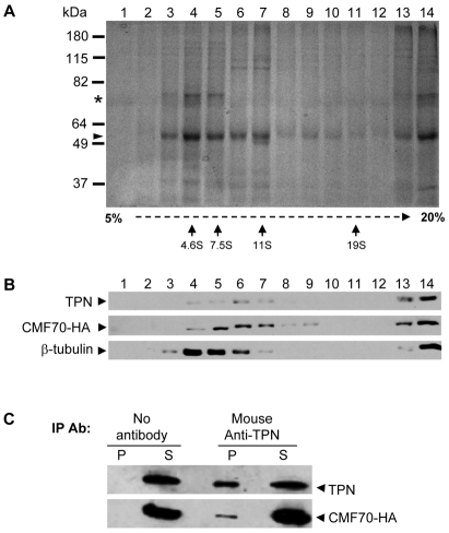
Fig. 3.
TbCMF70 is in a complex with trypanin. (A,B) Sucrose gradient fractionation of solubilized flagellar skeletons. Flagellar skeletons from CMF70-HA cells were extracted with 0.5 M KI, dialyzed against immunoprecipitation buffer and centrifuged to remove insoluble material. The soluble fraction was subjected to 5–20% sucrose gradient centrifugation and fractions were analyzed by SDS–PAGE and Coomassie Blue staining for total protein (A) or immunoblotting using anti-trypanin (TPN), anti-HA or anti-β-tubulin antibodies as indicated (B). In A, arrows indicate the position of migration in sucrose gradients for calibration standards. Tubulin (arrowhead) and the PFR1 and PFR2 (asterisk) proteins are indicated. (C) Solubilized flagellar skeletons from CMF70-HA T. brucei cells were immunoprecipitated with or without the mouse monoclonal anti-TPN antibody and analyzed by immunoblotting of the supernatants (S) and precipitated beads (P) using rabbit polyclonal anti-trypanin (TPN) or mouse monoclonal anti-HA antibodies (CMF70-HA).

