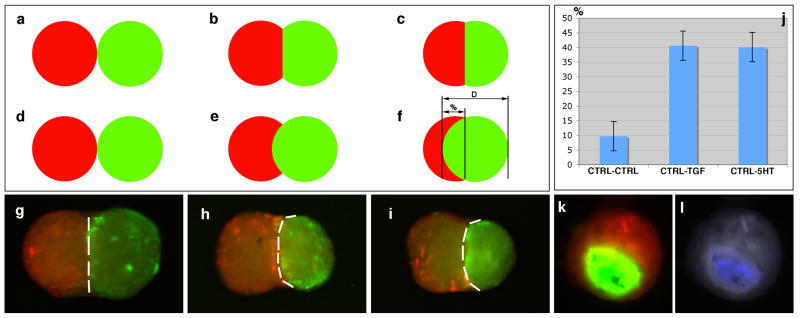
Figure 1.
Tissue spheroid envelopment assay using fluorescently labeled cells. a-f. Model quantitative assessment of spheroid envelopment. D = diameter, % = percent of envelopment. g-i. Tissue spheroids biofabricated from bone marrow cells after 72 hours exposure to control (g) medium, TGFβ1 (h), and serotonin (i). In panels h and i, control aggregates are red, while maturogen-treated aggregates are green. j. Percent envelopment of spheroids in response to maturogenic factors (TGFβ1, serotonin). k. Envelopment of spheroids biofabricated from cardiac valve interstitial cells. l. Immunodetection of HSP47 (a marker of collagen synthesis) in spheroids biofabricated from cardiac valve interstitial cells.

