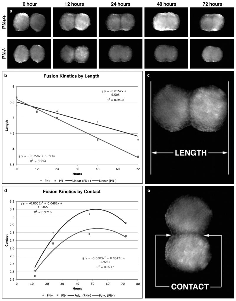
Figure 2.
Tissue spheroid fusion assay using wild-type and periostin-null dermal fibroblasts. a. Fusion of wild-type and PN-null spheroids biofabricated from dermal fibroblasts, imaged over 72-hours. b,c. Fusion kinetics by overall length (Blue = wild-type; red = PN-null). d, e. Fusion kinetics by contact region (Blue = wild-type; red = PN-null).

