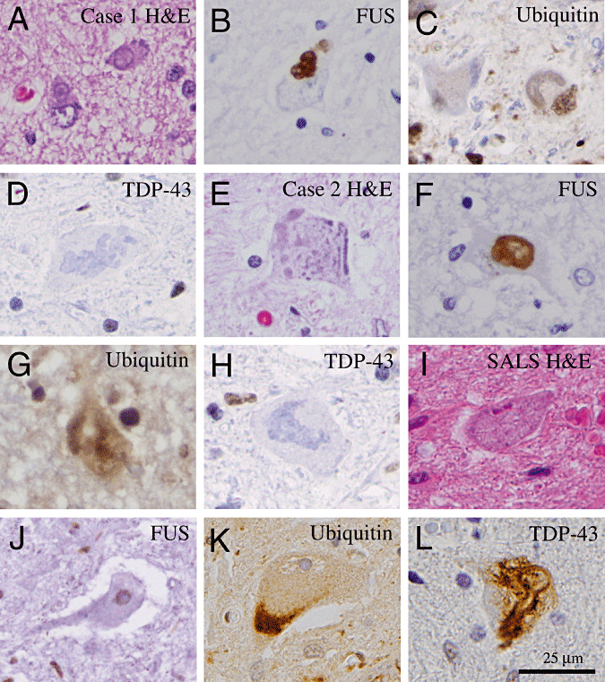
Figure 2.

Histopathology of juvenile amyotrophic lateral sclerosis (ALS) with basophilic inclusions in comparison with that of late‐onset sporadic ALS. (A,E) Hematoxylin and eosin (H&E) sections of the spinal cord show the presence of basophilic inclusions in the remaining motor neurons in Cases 1 and 2. (B,F) Immunohistochemical staining shows that the basophilic inclusions are positive for FUS proteins. (C,G) Furthermore, these inclusions are only weakly positive for ubiquitin but negative for TDP‐43 (D,H). (I) In contrast, spinal motor neurons in patients with late‐onset sporadic ALS show no evidence of basophilic inclusions on H&E‐stained section. (J) Furthermore, immunohistochemistry shows the presence of FUS protein in the nucleus of remaining spinal motor neurons. (K,L) Many of the spinal motor neurons in late‐onset sporadic ALS patients show abnormal accumulation of ubiquitinated proteins (K) and TDP‐43 proteins (L). Scale bar in L is 25 µm and applies to all panels.
